
Lung Segmentation Using ResUnet++ Powered by Variational Auto Encoder-Based Enhancement in Chest X-ray Images
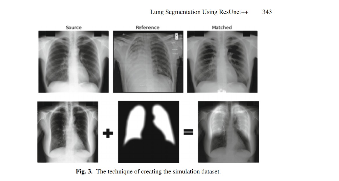
X-ray has a huge popularity around the world. This is due to its low cost and easy to access. Most of lung diseases are diagnosed using Chest X-ray (CXR). So, developing computer aided detection (CAD) provided with automatic lung segmentation can improve the efficiency of the detection and support the physicians to make a reliable decision at early stages. But when the input image has image artifacts, then any lung segmentation model will introduce suboptimal lung segmentation results. In this paper, a new approach is proposed to make the lung segmentation model robust and boost the basic models’ performance. This is done through two stages: image enhancement and lung segmentation. The first stage aims to enhance the image quality by using the combination of Variational Autoencoder (VAE) and U-Net. The features of VAE and U-Net are concatenated to decode the enhanced output CXR image. The second stage is segmenting the lung using ResUNet++, which can perform well with a few number of images. Moreover, it combines the advantages of residual blocks, squeeze and excitation blocks, Atrous Spatial Pyramidal Pooling (ASPP), and attention blocks. The proposed method is trained on JSRT dataset. The proposed approach achieved Dice score of 0.9848, 0.99 and the Jaccard score of 0.9783, 0.984 on test data of NLM-MC and JSRT datasets, respectively, which outperforms results in other state of the art models. © 2022, The Author(s), under exclusive license to Springer Nature Switzerland AG.
Related Publications
Hands-on analysis of using large language models for the auto evaluation of programming assignments